
Spotlight: Buffy Coat
What is Buffy Coat?
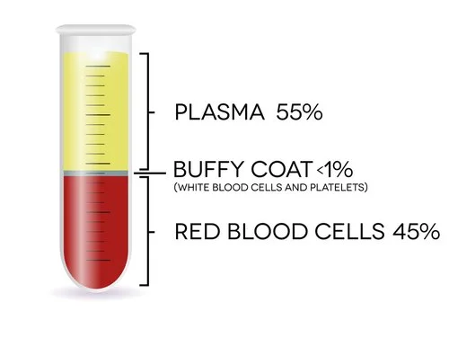
The buffy coat is a component of human blood that contains a high concentration of white blood cells (leukocytes) and platelets. It is typically found between the red blood cell layer (at the bottom of the blood sample) and the plasma layer (at the top) after blood has been centrifuged. The buffy coat appears as a thin, whitish layer and is composed of various types of white blood cells, including lymphocytes, monocytes, and granulocytes, as well as platelets.
How do you isolate buffy coat?
The collection of the buffy coat involves a process called centrifugation, which separates the different components of whole blood based on their density. After a blood sample is drawn from a donor or patient, it is placed in a centrifuge and spun at high speeds. This causes the heavier red blood cells to settle at the bottom of the tube, while the lighter plasma rises to the top. The buffy coat, which is denser than plasma but lighter than red blood cells, forms as a distinct layer between the two.
Once the blood has been centrifuged, the buffy coat can be carefully aspirated or pipetted from the top of the red blood cell layer. It is then processed further for various applications, such as isolating specific cell types for research or clinical purposes. Buffy coat samples can be used to obtain purified populations of white blood cells for immune cell profiling, genomic analysis, or functional studies. Additionally, the platelet-rich fraction of the buffy coat can be isolated and used in platelet transfusions or research related to hemostasis and thrombosis.
How Buffy Coat revolutionized cancer research
The revolutionization of cancer research by the buffy coat lies in its potential as a rich source of tumor cells, circulating tumor DNA (ctDNA), and other biomarkers relevant to cancer diagnosis, prognosis, and treatment monitoring. By isolating and analyzing circulating tumor cells (CTCs) or ctDNA from the buffy coat, researchers can obtain valuable insights into cancer biology, tumor heterogeneity, and treatment response. This non-invasive approach, known as liquid biopsy, offers several advantages over traditional tissue biopsy, including the ability to capture tumor dynamics in real-time, assess treatment resistance, and monitor minimal residual disease.
Liquid biopsy using buffy coat samples has revolutionized cancer research by enabling early detection of cancer, prediction of treatment response, and identification of therapeutic targets. By analyzing genetic mutations, epigenetic alterations, and other molecular markers in ctDNA or CTCs isolated from the buffy coat, researchers can tailor personalized treatment strategies for cancer patients, leading to improved clinical outcomes and quality of life. Moreover, liquid biopsy using buffy coat samples has the potential to overcome the limitations of tissue biopsy, such as tissue availability, invasiveness, and sampling bias, making it a valuable tool in precision oncology and translational research.